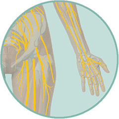
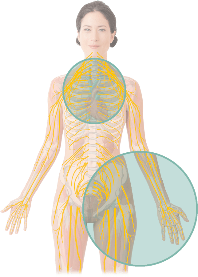
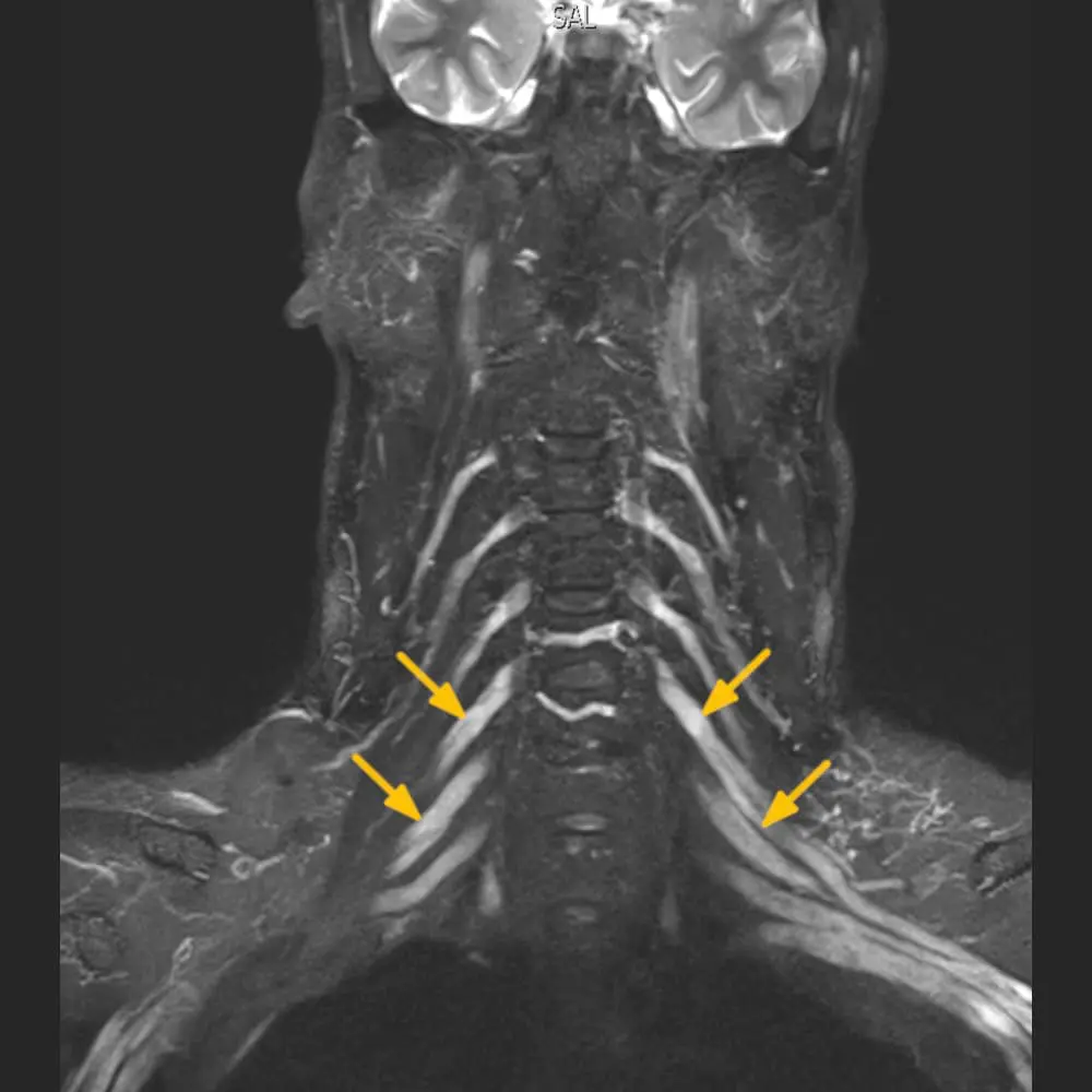
With MR neurography (also Nerve MRI), our radiologists examine affected regions of the body specifically. For example, the nerves can be visualised starting from the upper spine/neck via the arm-nerve plexus and the arm down to the fingers. In addition, we also examine the neighbouring anatomical structures such as joints, bones and muscles.
Pain can occur in very different ways. For example, it can "pull" or "shoot" from one region to another, such as from the lower back to the toes. However, it can also be limited to individual areas of the body and occur only at rest or only with exertion.
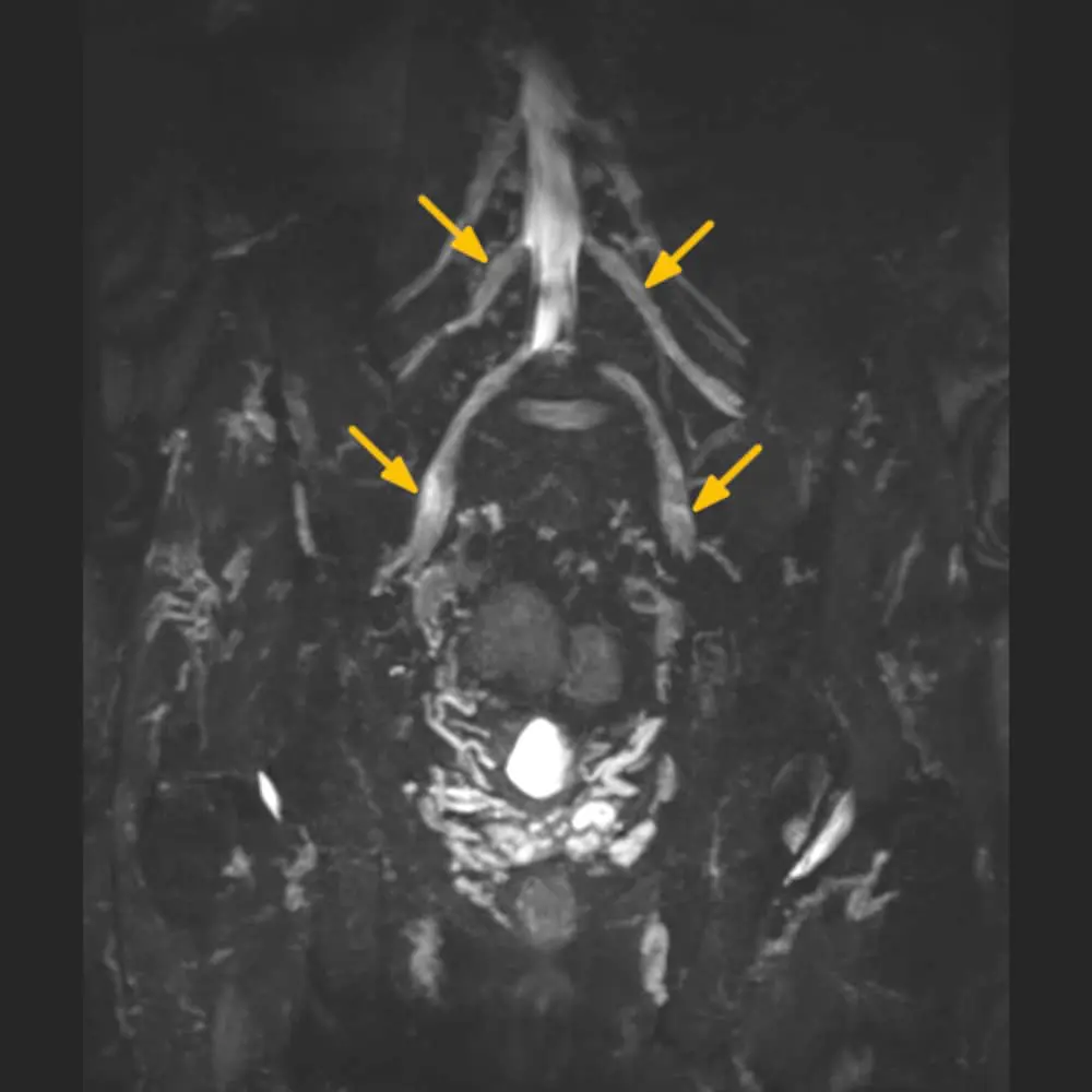
MR neurography is often the best examination method for initiating a suitable therapy to eliminate or alleviate your pain. The nerve MRI can be used to visualise longer sections of nerves precisely. With this scan, our experts can assess a nerve's condition down to the nerve bundle's level. In addition, we can clarify alternative causes for the respective pain.
Our peripheral nerve experts plan the MR examination carefully according to your pain history. Our lab technicians will scan you in the supine position, which usually takes about 45 minutes.
If necessary, we can also carry out targeted pain therapies under direct control in the MR scanner or ultrasound. In addition, ARISTRA offers counselling and second opinions for diseases of the nervous system.
The MRI examinations of the nerves at ARISTRA are continuously developed in cooperation with internationally recognised experts in neurological imaging. This means the images can be taken on-site using the most modern imaging technology and the latest scientific knowledge. Our neuroradiology experts evaluate your images.