MRI examination of the abdomen – clarifying diseases

Complaints in and around the abdominal cavity can severely restrict the everyday life of those affected. No matter the symptoms, an MRI examination can clarify whether there are changes in the various anatomical structures in the abdominal cavity. In this way, imaging employing abdominal MRI help make a decisive contribution to the diagnosis of diseases – completely without invasive surgical intervention.
Table of contents
MRI appointments at short notice
Findings shortly after the examination
State-of-the-art technology at the cutting edge of science
radiation-free examination

When to have an abdominal MRI?

Abdominal MRI at ARISTRA Illustration
An MRI examination of the abdomen is helpful for many different complaints. These include digestive problems and acute and chronic pain in the pelvis. Typical diseases can be, for example:
  • Inflammatory bowel diseases such as Crohn's disease or ulcerative colitis.
  • Inflammations of other organs, such as the liver, gall bladder, pancreas and more
  • Benign and malignant tumours
  • Gallstones or kidney stones
  • Abdominal wall hernias, inguinal hernias
An abdominal MRI examines a large area and provides an initial, often decisive, indication of the cause of the complaints. Depending on the individual situation, a targeted further examination of, for example, a single organ such as the liver, the kidney or the small or large intestine may be helpful.

The procedure of an abdominal MRI at ARISTRA

An abdominal MRI is performed comfortably in the supine position and lasts between 20 and 60 minutes, depending on the problem. During the treatment, you will wear hearing protection, as the strong magnetic field in the MRI machine causes loud knocking noises. The patient is usually pushed head-first into the MRI machine. At most of our ARISTRA MRI locations, we use MRI machines with an opening of 70 centimetres. The large opening means that an examination is usually possible without any problems. However, we always have a sedative on hand for anxious patients to make the scan possible.

Good to know: Unlike a CT scan, an MRI scan is radiation-free and, therefore, less stressful for the body.

MRI procedure - what you need to know!

Do I have to be sober for the abdominal MRI?
Depending on the exact areas our radiologists examine, it may be necessary to be sober before an MRI of the abdomen. Fasting is especially true before an examination of the bile ducts (also called MRCP) and before a scan of the gastrointestinal tract. However, before the study, we inform our patients about the preparation before the abdominal MRI.
MRI of the abdomen and contrast medium
During the MRI examination, a contrast agent will be injected via the arm vein. The agent accumulates differently in different tissue types, enabling our radiologists to get a detailed view of the internal organs. Contrast medium is used in particular when tumours or inflammatory diseases are being investigated. At ARISTRA, we use only tried and tested, safe contrast media at all locations, which is excreted within one day without any problems if the kidneys function normally.

MRI of the abdomen – what do you see?

Doctors divide the abdominal cavity into three areas. Radiologists can examine the entire abdominal cavity or only parts of the abdomen during an MRI. It is essential to know that complaints in the middle abdomen can also originate in the upper abdomen and vice versa. A comprehensive examination is vital for a precise diagnosis if there is no initial suspicion of the causes of the pain.
MRI upper abdomen – stomach, liver & co.
The upper abdomen is the area between the ribs and the belly button. Complaints in this area can have many different causes. Hence, a comprehensive MRI examination helps find a clue and helps patients quickly. The following organs belong to the upper abdomen:
  • Stomach
  • Liver
  • Gall bladder
  • Pancreas
  • Spleen
  • Intestine
  • Other sections of the small and large intestine
Some of these areas can be examined as part of a separate MRI – for example, the pancreas or the liver.
MRI examination of the middle and lower abdomen
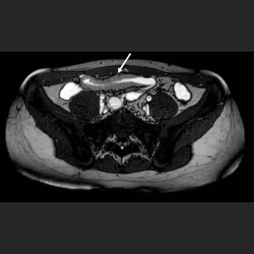
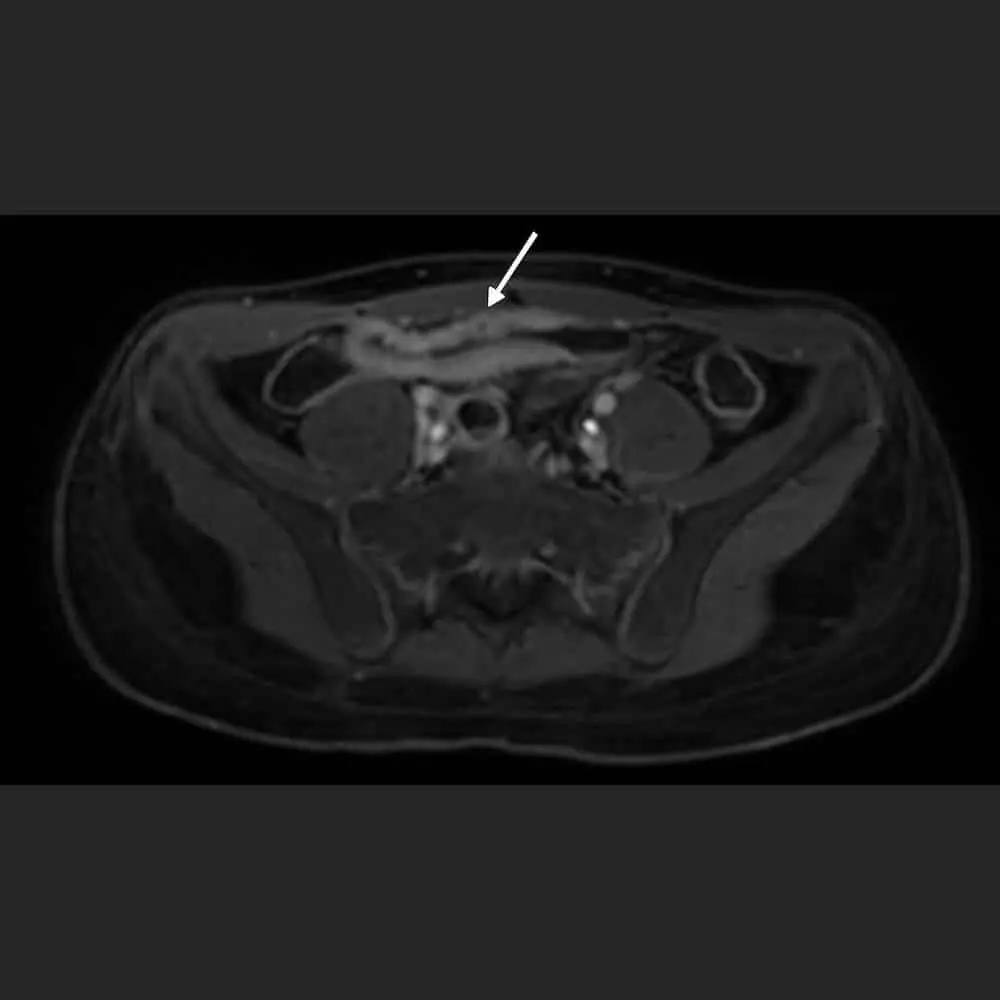
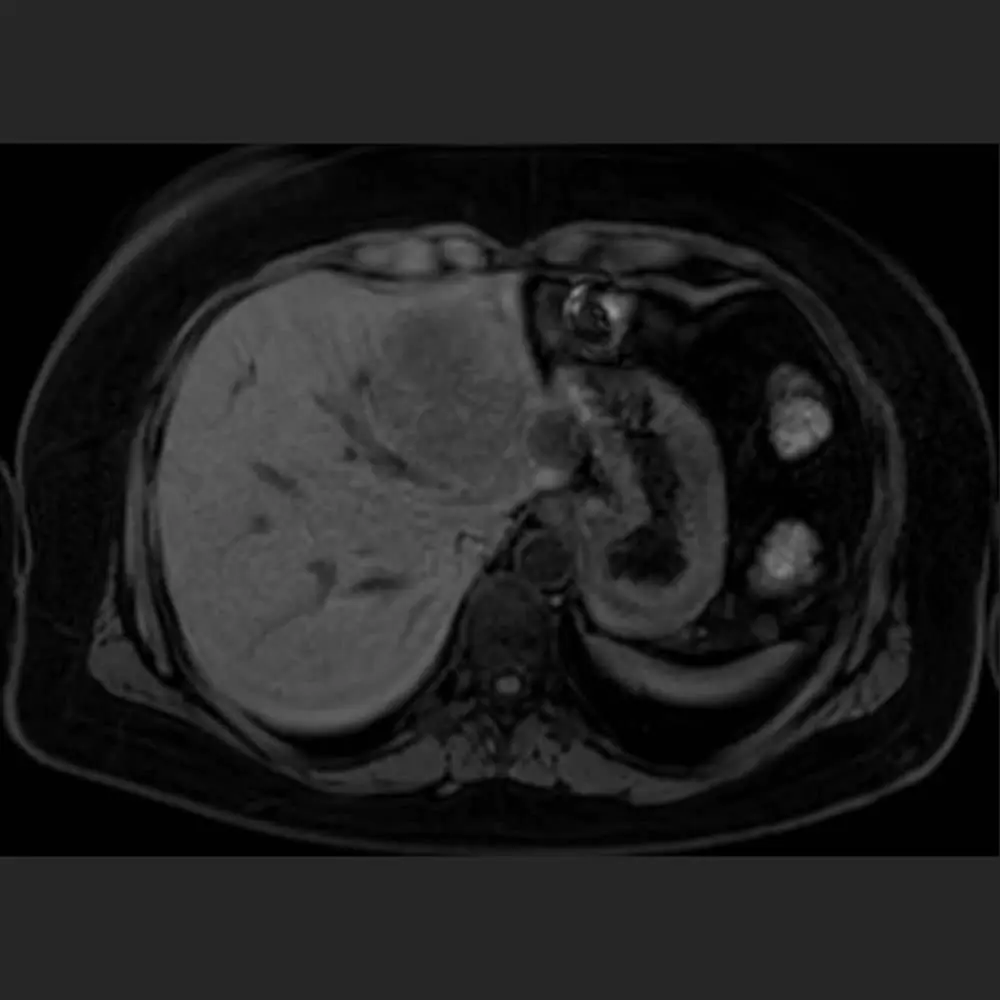
The mid-abdomen is not precisely delineated and covers approximately the area around the navel. Together with the lower abdomen, it includes the large and small intestines, the appendix, the kidneys, the pelvic viscera, and the urinary bladder. Suppose there is a suspicion of inflammatory processes in and around the intestine. In that case, radiology usually focuses on this area during MRI.

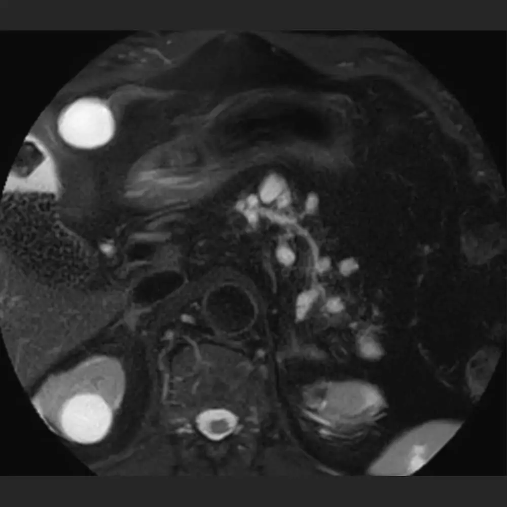
In women, the lower abdomen includes the uterus with fallopian tubes and ovaries. If there are complaints in this area, a gynaecologist will typically prescribe a separate MRI of the lower abdomen to detect, for example, inflammatory changes, endometriosis or gynaecological tumours.

In men, the prostate is part of the lower abdomen. An examination of the prostate is particularly important for cancer prevention. The prostate MRI is a more comfortable and precise diagnostic procedure for patients than the classic palpation examination.

Abdominal MRI » At a glance

The ARISTRA specialists look forward to welcoming you on-site. A pleasant atmosphere, short waiting times and personal medical care during the examination await you in the modern practice rooms.
Duration and price
approx. 30 minutes
Upon request*
Examination
Radiation-free
Quick appointment
* Prices depend on the individual case. Furthermore, we must consider your insurance status and whether you are a national or international customer. In most locations in Germany, we can only work with patients who have private insurance or are self-paying.

MRCP Abdomen MRI & MR Enterography (Sellink)

Magnetic resonance cholangiopancreatography (MRCP) is a particular form of abdominal MRI. This examination aims to visualise the gallbladder, the bile ducts and the ducts of the pancreas.
As a rule, a complete upper abdominal MRI is also carried out to ensure comprehensive clarification of the liver and pancreas. In this examination, a liver-specific contrast medium is generally used, enabling an optimised clarification of liver diseases. The liver-specific contrast medium is not usually administered in a general abdominal MRI.

Sellink MRI (MR enterography) examines the small intestine. Special preparation is necessary before the MRI. Patients should eat a low-fibre diet a few days before the examination. On the day of the scan itself, it is essential that you do not eat any solid food for 6 hours before the MRI.

In practice, you will be given a particular sugar solution to drink, ensuring that the small intestine is visible precisely in the images later. In addition, a contrast medium will be injected, and the intestinal activity will be briefly inhibited with another medication (e.g. Buscopan). These measures ensure that the MRI machine can produce sharp images, which our radiologists can evaluate well. The inhibited intestinal activity only lasts for a short time. The patients excrete the sugar solution and the contrast medium without problems during the day.

Advantages of the abdominal MRI

Magnetic resonance imaging of the abdomen offers a gentle and non-invasive examination for various complaints. Changes in the organs, such as inflammations, tumours and much more, can be examined uncomplicated. An MRI of the abdomen can be beneficial before an operation to get an idea of the abdominal cavity in advance.

In contrast to other examinations such as CT or X-ray examinations, MRI is radiation-free and can be used in most cases without any problems.

Abdominal MRI Images

Book your appointment with ARISTRA

Make an MRI appointment

Call us directly at
Alternatively, you can use our convenient online form. We will get back to you as soon as possible. We can usually offer you an appointment for your MRI at short notice.
EN Untersuchungen

To cancel an appointment, please use our appointment cancellation form.