MRI of Brain, Head, and Skull at ARISTRA

The brain is the centre of our central nervous system and a vital organ. Changes in this area often start subtly but can have serious consequences. An MRI (magnetic resonance imaging) of the brain, head, and skull can provide information about various diseases. As a patient at ARISTRA, you are in good hands. Our on-site radiologists and the international network of specialists offer you a safe diagnosis and thus enable a quick start to therapy.
Table of contents

When is a brain MRI useful?

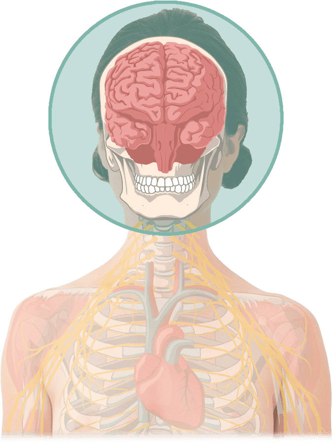
MRT Kopf & Gehirn bei ARISTRA Illustration
Magnetic resonance imaging of the head can be helpful for the following symptoms. It makes the different areas of the human brain visible and thus helps to detect or rule out diseases:
  • Persistent headaches and migraine
  • Feeling of pressure in the head
  • Facial pain
  • Recurrent dizziness
  • Tinnitus
  • Gait disturbances and other neurological / motor deficits
  • Speech problems
  • Forgetfulness
  • Balance disorders
  • Epilepsy
  • Multiple sclerosis
  • Sinus thrombosis
  • After a stroke
  • With cerebral haemorrhages
  • After severe head injuries
  • Trauma sequelae
  • Family history of related diseases
  • Developmental disorders in children

What areas does the radiologist see?

The imaging procedure enables a clear view of various structures in the head. A cranial MRI is the right way to clarify the causes of many different complaints. The following areas can be examined in radiology by an MRI scan:
  • Cerebral ventricles
  • Skull (skeletal part)
  • Teeth & Jaws
  • Eyes
  • Sinuses
  • Ears
  • Sustaining arteries

Procedure for an MRI examination of the brain

Prof. Dr. med. Philipp Bäumer

Certified specialist for radiology and neuroradiology
Expert for Neuroradiology
Prof. Dr. med. Philipp Bäumer is a specialist in radiology with an additional specialisation in neuroradiology. He completed his habilitation in neuroradiology at the University of Heidelberg, where he acquired great expertise in the imaging of peripheral nerves over many years.
Prof. Dr. med. Philipp Bäumer is a specialist in radiology with an additional qualification in neuroradiology. He is an expert in the imaging of peripheral nerves.
Prof. Dr. med. Wibke Müller-Forell

Certified specialist for radiology
Expert for the orbita
Prof. Dr. med. Wibke Müller-Forell is Professor Emeritus at the Johannes Gutenberg University in Mainz. There she was acting head of the Institute for Neuroradiology. Her clinical focus is on diseases of the orbita, MR diagnostics of the central nervous system, and interventional stroke therapy.
Prof. Wibke Müller-Forell is Professor Emeritus at the Johannes Gutenberg University Mainz, where she was acting head of the Institute for Neuroradiology.
PD Dr. med. Yasemin Tanyildizi

Certified radiology specialist
Expert for Neuroradiology
PD Dr. med. Yasemin Tanyildizi is a certified specialist in radiology. She is an expert in neuroradiology and did research and habilitation at the University Medical Centre Mainz for many years. She has also completed numerous stays abroad, including in Australia and Turkey. She is the author of significant international scientific publications.
PD Dr. med. Yasemin Tanyildizi is a certified specialist in radiology and an expert in neuroradiology. She researched at the University Medical Centre Mainz.
Dr. med. Kira Lutz
Dr. med. Kira Lutz

Certified radiology specialist
Expert for Neuroradiology
Dr. med. Kira Lutz has a medical specialization in radiology with a focus on neuroradiology. She has worked and researched at Heidelberg University Hospital and Freiburg University Hospital.
Dr. med. Kira Lutz has a medical specialization in radiology with a focus on neuroradiology. She researched at the Heidelberg University Hospital and in Freiburg.
PD Dr. med. Erika Jung-Ulbrich

Certified radiology specialist
Specialist musculoskeletal system
PD Dr. med. Erika Jung-Ulbrich is an expert in musculoskeletal radiology, with a focus on MRI of the musculoskeletal system. She completed her habilitation in musculoskeletal radiology at the University of Zurich and worked for several years as a senior physician at the University Hospital Zurich.
PD Dr. med. Erika Jung-Ulbrich is an expert in musculoskeletal radiology, with a focus on MRI of the musculoskeletal system.
Prof. Shinji Naganawa

Certified radiology specialist
Expert for inner ear imaging
Prof. Shinji Naganawa is a pioneer in inner ear imaging. He has researched the optimization of MR protocols for detecting internal ear pathologies for over 25 years. He is clinically active as Director of the Department of Radiology at Nagoya University Hospital, Japan.
Prof. Shinji Naganawa is a pioneer in inner ear imaging. He has researched the optimisation of MR protocols for internal ear pathologies for over 25 years.
Prof. Dr. med. Lorenz Jäger

Specialist in diagnostic radiology
Expert for oncological and ENT radiology
Prof. Dr. med. Lorenz Jäger is an expert in ENT radiology, neuroradiology, musculoskeletal radiology and oncological radiology and completed his specialist training at the LMU in Munich. He has held two visiting professorships since 2018.
Prof. Dr. med. Lorenz Jäger is an ENT, neuro-, musculoskeletal and oncological radiology expert. He has held two visiting professorships since 2018.
Dr. med. Andrea Godau

Specialist in diagnostic radiology and neuroradiology
Dr. med. Andrea Godau completed her training at Freiburg University Hospital. After subsequently working as a senior physician in Sweden, she acquired her specialisation in neuroradiology at the University Hospital Freiburg.
Dr. med. Andrea Godau completed her training at Freiburg University Hospital. She has a specialisation in neuroradiology.
Dr. med. Thomas Hambrock

Certified radiology specialist
Expert for prostate and oncological radiology
Dr. med. Thomas Hambrock is an expert in prostate cancer and oncological radiology. The focus of his research activities at Radboud University Hospital, Nijmegen, the Netherlands, is on the development of advanced MRI sequences in oncological imaging.
Dr. med. Thomas Hambrock is an expert in prostate cancer and oncological radiology. He is researching the development of oncological MRI sequences.
Dr. med. Michael Ho
Dr. med. Michael Ho

Certified radiology specialist
Expert for Peripheral Nerves
Dr. med. Michael Ho is a certified radiology specialist. He has worked clinically and scientifically on peripheral nerve imaging at the University Hospital Heidelberg. Most recently, he worked at University Hospital Zurich, among others, and is an expert in this field. He is the managing director and co-founder of ARISTRA.
Dr. med. Michael Ho is a certified radiology specialist. He is an expert in peripheral nerve imaging and the managing director and co-founder of ARISTRA.
Dr. med. Yvonne Böckenfeld

Certified specialist for ear, nose, and throat medicine
Expert for ENT
Dr. med. Yvonne Böckenfeld is a certified specialist in ear, nose, and throat medicine in Karlsruhe, Germany.
PD Dr. med. Johann-Martin Hempel

Certified radiology specialist
Expert in neuroradiology and head and neck radiology
PD Dr. med. Johann-Martin Hempel is an expert in neuroradiology and head and neck radiology. He practised for many years at the University Hospital of the Eberhard Karls University in Tübingen, where he completed his habilitation.
PD Dr. med. Johann-Martin Hempel is an expert in neuroradiology and head and neck radiology. He practised at the University Hospital in Tübingen for many years.
Dr. med. Matthias W. Wagner

Certified specialist for radiology and neuroradiology
Expert for pediatric neuroradiology
Dr. med. Matthias Wagner is an expert in pediatric neuroradiology. He gained his experience in Switzerland, the USA and Canada. For ARISTRA, he works as a teleradiologist for paediatric and adult neuroradiology.
Dr. med. Matthias Wagner is an expert in pediatric neuroradiology. He gained experience in Switzerland, the USA and Canada.
Dr. med. Kai-Thorsten Müller

Specialist for radiology
Expert in abdomen, neurology and orthopaedics
Dr. med. Kai-Thorsten Müller has many years of experience in cross-sectional imaging of the musculoskeletal system in neurology and the abdomen.
The MRI examination of the brain takes place in the supine position and lasts 20-30 minutes. The head must lie in the MRI machine - an additional coil must be placed over the head for some questions. At ARISTRA, we use MRI machines with an opening of 70 centimetres. Nevertheless, an MRI of the skull can be unpleasant for anxious patients. Therefore, we always keep a sedative at hand. You can also interrupt or cancel the examination anytime with an emergency button. Be aware that an interruption or cancellation can lead to the scan being unusable.

Before the MRI, you should remove all metal objects, such as jewellery, watches, and similar. It would help if you avoided make-up such as rouge, eye shadow or lipstick, as these often contain metallic components.
Good to know: A cranial MRI usually uses what is known as diffusion tensor imaging. It measures the diffusion movements of water molecules in the body and displays them. With this scan, you can reliably detect diseases of the central nervous system.
Immediately after the examination, the radiologists on site will discuss the findings with you. In addition, your referring doctor will also receive the results and plan further therapy steps together with you. The ARISTRA radiologists work with the ARISTRA network for complex questions and unclear findings. Here you will find international specialists in every field of radiology. In this way, patients always receive a comprehensive and precise diagnosis.

MRI contrast agent - when is it necessary?

A contrast medium is not always necessary for a brain MRI. However, always when vessels or inflammations are to be displayed more precisely during the examination. The contrast medium helps to visualize even the finest structures. This way, various diseases, such as inflammatory changes and other pathological processes, can be assessed very well. In contrast to many contrast agents used in CT (computed tomography), this is iodine-free. If you are allergic to certain contrast media or suffer from impaired kidney function (renal insufficiency), please state this before the examination. At ARISTRA, our radiologists use a contrast medium that has been tried and tested over many years. Healthy kidneys can break it down quickly in most cases.

Brain MRI during depression – the fMRI

With the help of a functional MRI (fMRI), changes in blood flow and metabolic processes in the brain can be made visible. The examination consists of a so-called pre-scan, an anatomical MRI image and the actual fMRI. As soon as one of our brain areas is activated - for example, because we have to think of something specific or solve a task - the metabolism in this area is ramped up. Our radiologists can follow these changes based on the MRI images.

The procedure is used, among other things, to diagnose depression. But fMRI is also used for epilepsy, Alzheimer's, and Parkinson's. In the case of depression, you must first rule out inflammatory, vascular or degenerative processes and a brain tumour.

As fMRI is a highly specialised procedure, we can only offer this examination at selected locations. Feel free to contact us; our staff will advise you.
MRI Brain » At a glance

The ARISTRA specialists look forward to welcoming you on-site. A pleasant atmosphere, short waiting times and personal medical care during the examination await you in the modern practice rooms.
Duration and price
approx. 30 minutes
Upon request*
Examination
Radiation-free
Quick appointment
* Prices depend on the individual case. Furthermore, we must consider your insurance status and whether you are a national or international customer. In most locations in Germany, we can only work with patients who have private insurance or are self-paying.

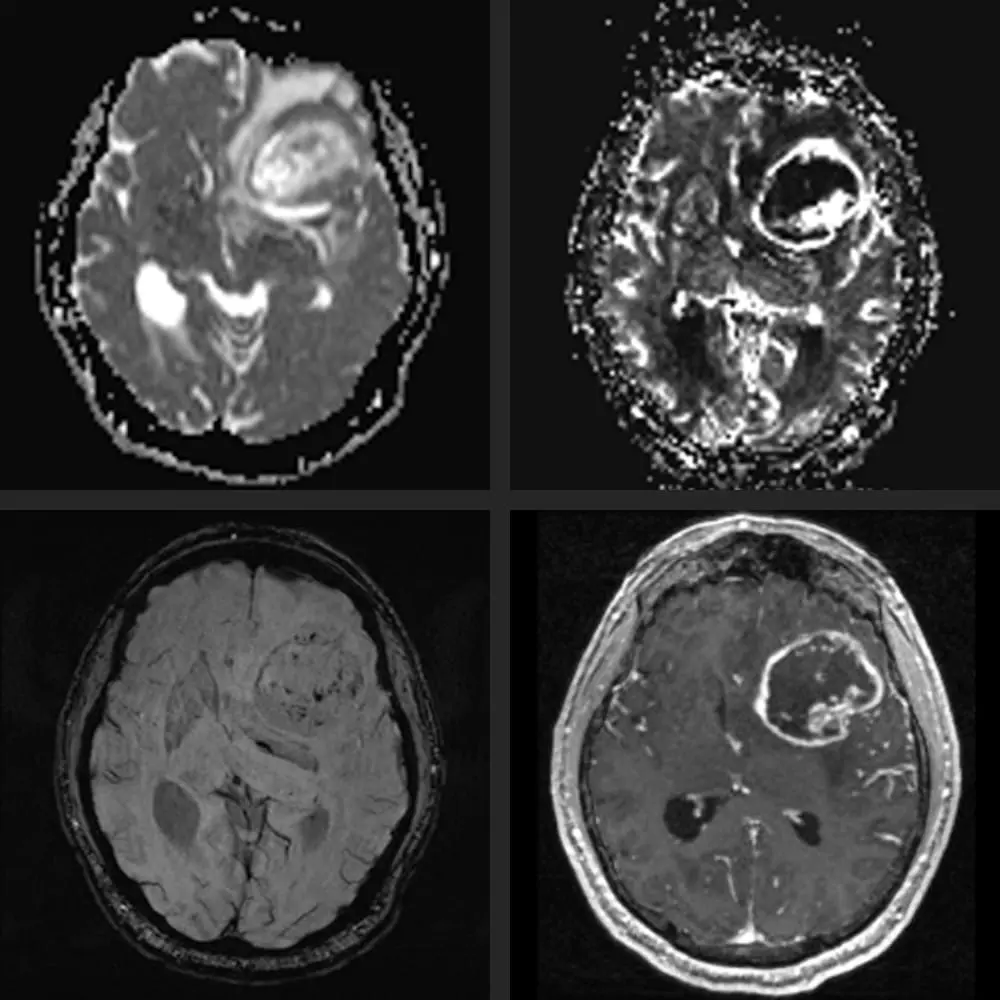
Brain tumour - reliable clarification in the MRI

Patients who repeatedly suffer from headaches often fear that they are suffering from a brain tumour. However, the risk of developing a brain tumour without a genetic predisposition is less than one per cent. An MRI is the safest and most accurate way to clarify the suspicion of a tumour in the brain. At ARISTRA, we carry out the scans thoroughly. Thanks to fast diagnostics, we can enable you to start therapy early or give you the all-clear quickly.

Detecting diseases of the brain, head, and skull on MRI

The following diseases can be reliably detected or excluded with the help of an MRI examination of the brain:
  • Epilepsy
  • Parkinson's disease
  • Dementia
  • Tumour in the cranial region or brain
  • Meningitis
  • Creutzfeld-Jacob
  • Circulatory disorders
  • Malformations and deformities
  • Developmental disorders
  • Multiple Sclerosis
  • Inflammations, constrictions & calcifications
  • Pathological vascular dilatations
  • Craniocerebral trauma
  • Metabolic disorders
  • Degenerative brain changes
  • Vascular aneurysm

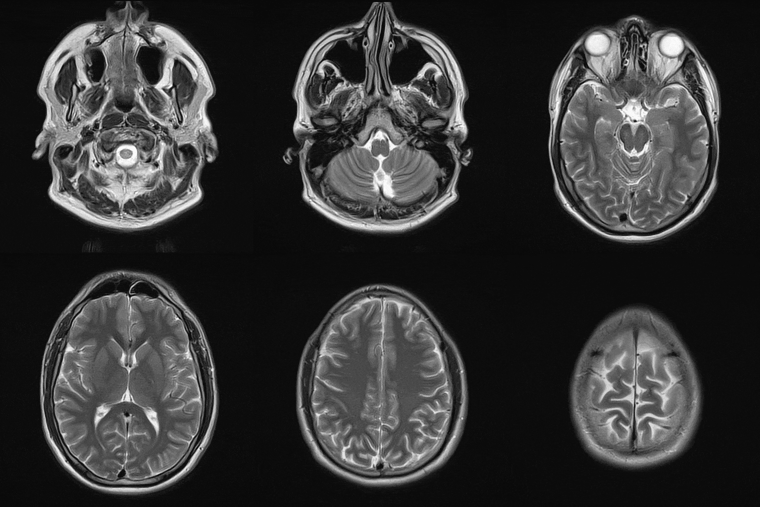
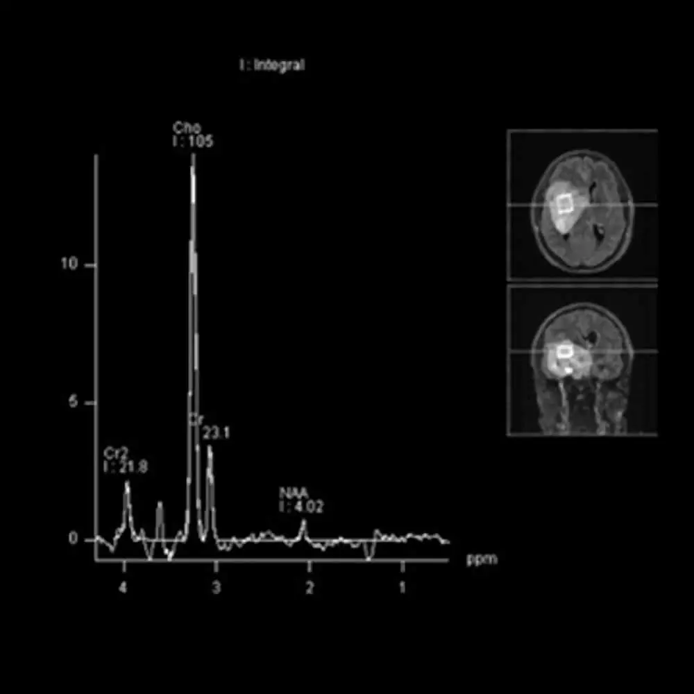
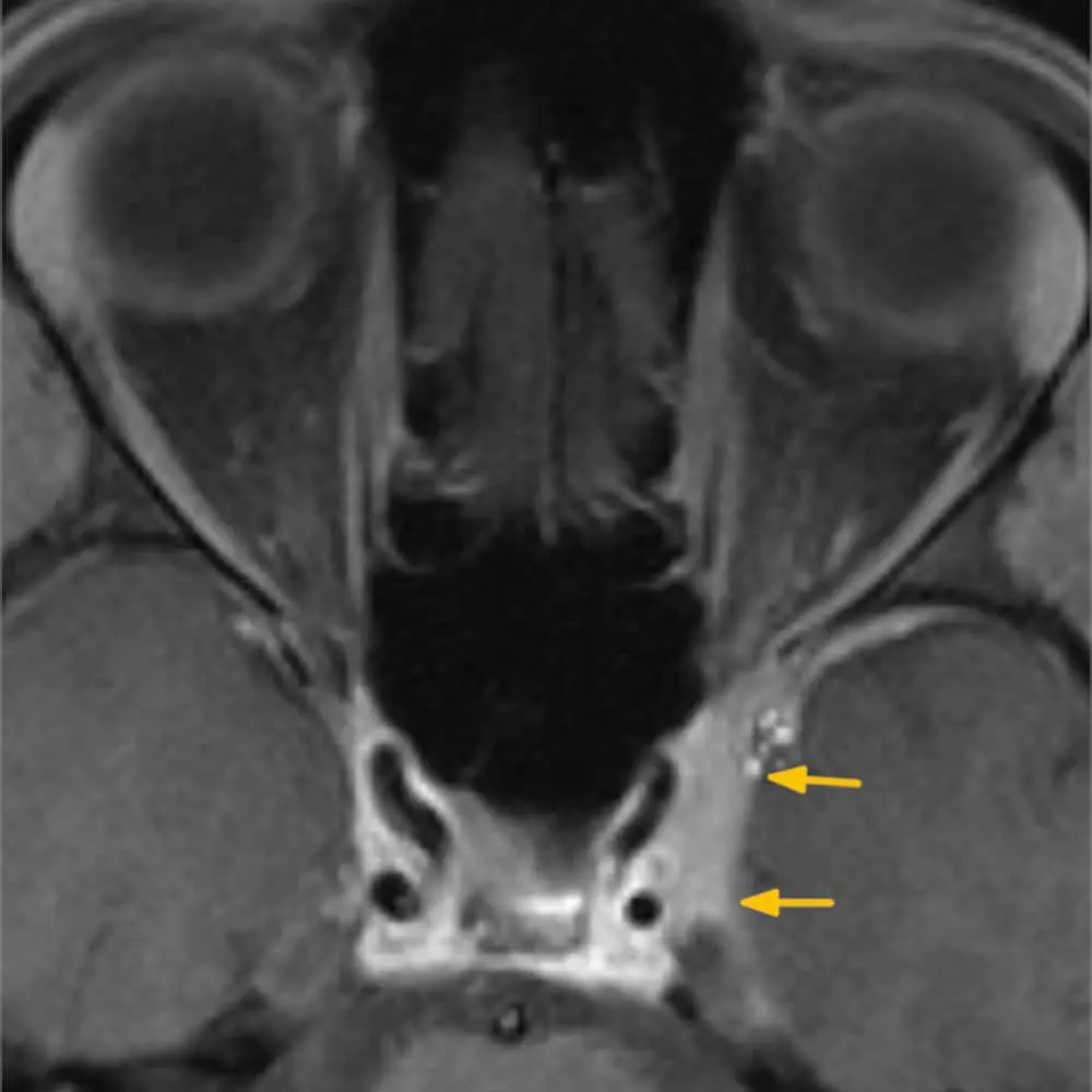
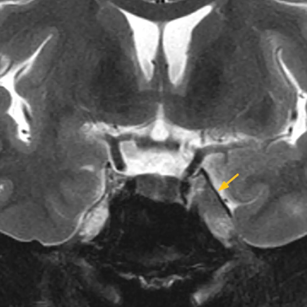
MRI Brain Images

Book your appointment with ARISTRA

Make an MRI appointment

We can usually offer you an appointment for an MRI examination at short notice. You can request your preferred appointment via this form.
EN Untersuchungen

To cancel an appointment, please use our appointment cancellation form.